New method of dental imaging uses squid ink for a painless, non-invasive check-up
09/18/2017 / By Jhoanna Robinson
According to engineers at the University of California San Diego, people who are getting checked at their dentists’ offices for gum disease need not worry anymore about pain and discomfort. Why? Because squid ink is the answer to their problems.
In a study that was published in the Journal of Dental Research, the researchers developed a new dental imaging method to look at a patient’s gums by combining cuttlefish ink with light and ultrasound.
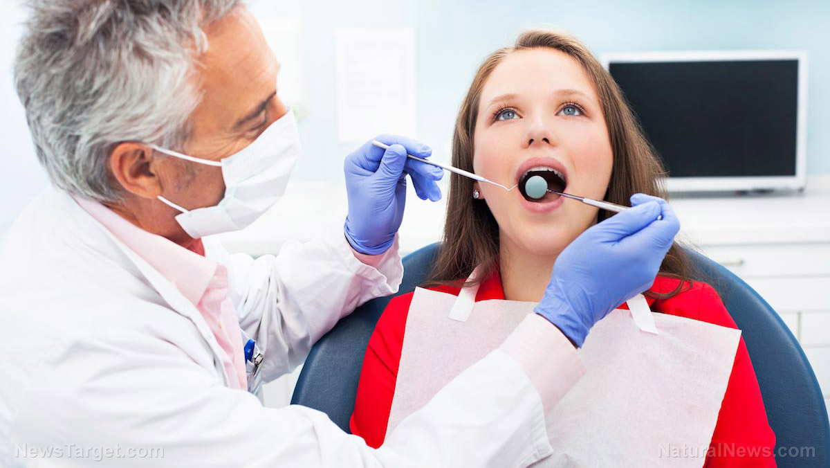
Traditionally, dentists assess the condition of their patients’ gums using an instrument called periodontal probe, which is a thin, hook-like metal tool that has labels like a measuring stick and is placed in between the teeth and gums to see how much the gums have shrunk back from the teeth, causing the formation of pockets.
A pocket depth that measures one to two millimeters shows healthy gums while a pocket depth of three mm is indicative of gum disease. The deeper the pockets, the worse the gum disease. (Related: Contrary to what your dentist may tell you, you CAN reverse tooth decay, new research finds.)
However, the researchers mentioned that dentists often measure pockets differently, and that the probhe can only measure the pocket depth of one spot at a time.
The researchers said that the process of identifying gum disease via squid ink begins by rinsing the mouth with a paste that is made of food-based squid ink mixed with water and cornstarch. The squid ink then would cling to the pockets in between the gums and the teeth. Next, an ultrasound would be taken of the patient’s mouth.
The squid ink-based rinse acts as a contrast agent for an imaging method called photoacoustic ultrasound. This involves shining a white, laser pulse onto a sample, which heats up and expands, giving off an acoustic signal that researchers can examine.
“Using the periodontal probe is like examining a dark room with just a flashlight and you can only see one area at a time. With our method, it’s like flipping on all the light switches so you can see the entire room all at once,” senior study author and UC San Diego nanoengineering professor Jesse Jokerst said.
This technique makes it possible for dentists to visualize a full map of the pocket depth around each tooth – a remarkable improvement over the traditional method.
Researchers said the results of the photoacoustic imaging method were consistent across multiple tests, unlike when the assessment was done with the periodontal probe, wherein results varied from one test to another.
Jokerst said the idea of using squid ink started while he and some colleagues were having dinner in Tokyo. They brainstormed about possible contrast agents that wouldn’t be rejected by the United States Food and Drug Administration (FDA).
“The idea that a food could be used was intriguing, but which foods are dark and could potentially be used for imaging? Squid ink is perfect because it is a liquid and thus can penetrate into the space between the tooth and the gum, i.e., the pocket,” Jokerst said.
Squid ink is a good option, Jokerst said, since because it was so dark, it can readily absorb light. Plus, it is already edible.
“Getting materials FDA-approved is very time-consuming. We didn’t want to create a new material that would be subjected to years of scrutiny and testing. By using a material that is already approved as a food, we hope to speed up the time needed to get this into dental offices,” Jokerst concluded.
Read more stories such as this one at Dentistry.news.
Sources include:
Tagged Under: dental imaging method, dentistry, gum disease, natural health, oral health, periodontal probe, squid ink